

Diabetic Retinopathy
What can happen to my vision?
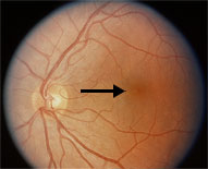
Normal Macula
The retina is a marvelous sensory tissue which gives us the gift of sight. This tissue is very sensitive to changes in structure and form. Areas of retina which are devoid of oxygen and nutrients atrophy and will no longer provide vision. Likewise, areas of retina which are swollen due to leakage of blood vessels will not provide vision. The area of retina which is most precious and provides us with our central or straight-ahead vision is known as the macula. This is the area of the highest density of sensory cells known as cones. If there is any disruption in this particular area our vision is greatly disturbed. This is the area of retina where macular degeneration occurs. The greatest threat to our vision occurs if there is leakage from the blood vessels in or in close proximity to this region of tissue. If swelling does occur in this area vision decreases dramatically, very quickly. Patients with swelling in this area will report distorted vision, such as a straight telephone pole, now appears crooked. They will also report that their central vision or acuity is greatly diminished. Such an event in this area may come suddenly as a blood vessel ruptures, or slowly and almost unnoticed with slow swelling taking place over time. A simple, self-monitoring tool is available to check this area for any changes. This self-monitoring tool is a small chart with grid lines known as the Amsler Grid. Any disturbance of the retinal tissue in this area will be detected by the patient on this chart. Patients at risk for developing swelling in this area should us this self-monitoring technique. One may test their vision in the privacy of their home as often as their eye doctor advises.
Another, more sophisticated in-office test that evaluates this area of retina is called a visual field exam. This test is done by an instrument that presents stimuli to the macula area of the retina in the form of small dots of light. These lights are presented to one eye randomly with differing intensity to determine if there is any loss of sensitivity of the sensory cells due any change in structure or function of the tissue. A new generation state-of-the-art visual field instrument was placed on the market about nine months ago to evaluate just this small, special area of retina. What sets this instrument apart from other visual field instruments is that it's calibration is based upon the earliest perceivable difference in light stimuli. This internal calibration data is from actual patients as they have encountered the earliest onset of pathological changes. These pathological changes in the macula area may be a result of natural degenerative tissue changes or changes in the tissue from the first sign of swelling.