

Diabetic Retinopathy
How & why may these changes occur in my retina?
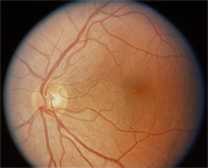
Normal Retina
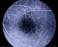
Fluorescein Angiography of Normal Retina
The retina is the sensory tissue on the back of the eye which absorbs light energy as Einstein describes as photons and transforms light energy into electrical impulses which are sent to our brain by way of the optic nerve. This is essentially the process of vision. The retina is a very complex tissue being only as thick as a credit card yet having ten distinct layers, each having a special job to perform. The retina miraculously performs this transisition of one form of energy to another. Because of this, many regard the retina as an extension of brain tissue.
The blood vessels which serve the retina lie in the middle of the retina. There are special protector cells on the surface of the blood vessels called pericytes. When blood sugar levels in our bloodstream remain elevated for prolonged periods of time or fluxuate greatly theses special protector cells die. This causes the walls of the blood vessels to weaken creating a microaneurysm. If the microaneurysm bursts blood leaks out into the retina. This extra fluid causes swelling within the retina, damaging the photoreceptor cells (rods and cones) consequently interrupting the process of vision. This interruption of vision is confined to the area of bleeding and swelling. Another problem that may occur with damaged blood vessels is that the vessels may collapse.
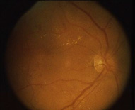
Abnormal retinal circulation system.
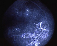
Note areas of leakage shown by fluorescein dye.
This collapse of blood vessels ceases blood flow to that area of retina. This area of retina has no oxygen, or becomes hypoxic and now atrophies due to lack of blood flow. These areas of atrophy are classified as hard exudates and cotton wool spots and are characterized by yellow areas against the normal red background of the retina. The doctor can easily recognize these areas of degenerated retina. These particular abnormalities in the retina are permanent in nature and vision is lost forever. The response of the retina to this hypoxic area is to grow new blood vessels to serve this oxygen-starved tissue. This process is called neovascularization. One may think this process is a noble effort by the retina but the problem with these new blood vessels created is they are only a single cell in thickness and may rupture very easily now causing a general hemorrhage in the middle of the eye known as the vitreous.
Damage to the blood vessels causing either leakage of blood into the retina or closure of blood vessels is the reason diabetic retinopathy develops. This all relates back to high, sustained levels of sugar in the bloodstream.
Retinal eye disease is silent. Changes in the retina may be taking place without the knowledge of anyone, and then all of a sudden one discovers through experience that there is a small problem or a large problem. We never desire to awaken one morning and reach this discovery point as it may be too late to save precious vision. Rather, the safe bet is to have regular eye exams to be alerted to potential problems and avoid any vision loss. Patients with diabetes are encouraged to have an eye exam annually, unless advised by your eye doctor otherwise.